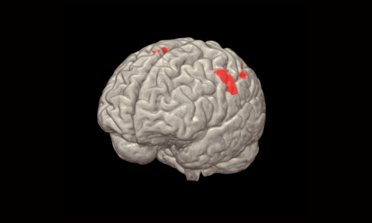

Functional magnetic resonance imaging (fMRI) is an advanced resonance technique (MRI) through which it is possible to identify the brain areas used (in jargon, “which are activated”) during the execution of a motor task, of language, visual or of another type. The fMRI exam is performed by asking the subject to perform the task (task) during the normal course of the exam. For example, the subject will be asked to move his fingers or grimace with his mouth. The areas that are activated are then shown in a color scale superimposed on normal morphological resonance images (see Figure)
The studies of functional MRI currently have a role of primary importance in scientific and clinical routine. In the research they are mainly used for the characterization and for the evaluation of anomalies of the main cerebral functional networks. In clinical practice they are predominantly used for pre-surgical planning, aimed at identifying and saving the main elusive areas whose removal would have an important social impact on the patient’s life.
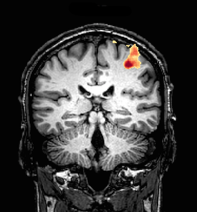
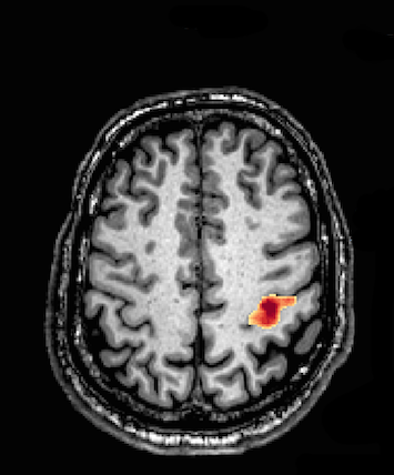
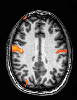
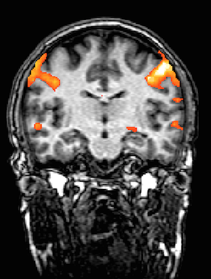
Figure: the first line shows an example of activation of the motor area (in a red-yellow scale) following a task during which the subject was asked to move his fingers (finger tapping). The second line activates after performing a series of grimaces with the mouth